BBYCT-131 Solved Assignment
- a) Give a brief account on lytic or lysogenic replication in viruses with suitable diagrams.
b) Discuss the role of bryophytes as ecological indicators - a) Define heterospory. What is its biological significance?
b) List the mechanisms of genetic exchange in bacteria and describe any one of them in detail. - a) Trace the development of female gametophytes in Cycas.
b) Enumerate the medical value of Gymnosperms. - Differentiate between the following pairs of terms:
i) Bacterial cell and Archaeal cell
ii) Liverworts and Mosses
iii) Transformation and Transduction
iv) Petrifaction and Compression - Prepare clear and well-labelled diagrams of the following:
i) Clamp connection formation in Basidiomycetes.
ii) V.S. thallus through a gemma cup in Marchantia.
iii) T.S. needle of Pinus.
iv) L.S. microsporangium of Selaginella. - a) Describe the internal and external structure of a typical bacterium.
b) Differentiate a bacterial cell from an archaeal cell. - a) Discuss different types of steles found in pteridophytes along with suitable diagrams.
b) Briefly describe the economic importance of Pteriodophytes or Gynosperms. - Prepare a detailed account of occurrence, morphology and ultrastructure in Cyanophyta.
- Define the transformation. How Griffith’s experiment was carried out to discover bacterial transformation?
- Write notes on the following:
i) Biofertilizer
ii) Polyembryony in gymnosperms
iii) Alternation of generations
iv) Coralloid roots of Cycas
Answer:
Question:-1(a)
Give a brief account on lytic or lysogenic replication in viruses with suitable diagrams.
Answer:
Lytic and Lysogenic Replication in Viruses
Viruses replicate through two primary cycles: lytic and lysogenic.
1. Lytic Cycle:
The lytic cycle is the process where a virus infects a host cell, hijacks its machinery to produce new virus particles, and causes the host cell to burst (lyse), releasing new viruses. The stages of the lytic cycle are as follows:
Stages of the Lytic Cycle:
- Attachment: The virus attaches to the surface of the host cell through specific receptors.
- Penetration: The viral genome is injected into the host cell, or the virus enters the host cell through endocytosis.
- Biosynthesis: The host cell’s machinery is hijacked to replicate the viral genome and synthesize viral proteins.
- Maturation: New viral particles (virions) are assembled from the replicated genome and synthesized proteins.
- Lysis: The host cell bursts (lyses), releasing the new virions into the environment, where they can infect new cells.
Diagram of the Lytic Cycle:

2. Lysogenic Cycle:
In the lysogenic cycle, the viral DNA integrates into the host cell’s genome, where it remains dormant for an extended period. The viral genome (provirus) can remain hidden within the host’s DNA and be passed on when the host cell divides. The cycle can switch to the lytic cycle under certain conditions, such as stress or environmental changes.
Stages of the Lysogenic Cycle:
- Attachment: The virus attaches to the host cell and injects its genetic material.
- Integration: The viral DNA integrates into the host cell’s chromosome, forming a provirus. The virus remains dormant and does not immediately cause cell lysis.
- Replication: The host cell divides, replicating the viral DNA along with its own. The viral genome is passed on to daughter cells.
- Induction: Under certain stress conditions, the provirus may become active and enter the lytic cycle.
Diagram of the Lysogenic Cycle:

Question:-1(b)
Discuss the role of bryophytes as ecological indicators.
Answer:
Role of Bryophytes as Ecological Indicators
Bryophytes, a group of non-vascular plants that includes mosses, liverworts, and hornworts, play a crucial role in ecosystem functioning and have been recognized as important ecological indicators. These plants, due to their sensitivity to environmental factors, can offer valuable insights into the health and stability of ecosystems, particularly regarding moisture, pollution, and habitat changes.
1. Sensitivity to Environmental Conditions
Bryophytes are highly sensitive to their surrounding environment because they lack vascular tissue, which means they do not have specialized structures for transporting water and nutrients. This makes them reliant on ambient humidity and atmospheric moisture. Consequently, they respond quickly to changes in temperature, humidity, light, and atmospheric pollutants. Their ability to reflect changes in these environmental factors makes them excellent indicators of ecological conditions.
2. Monitoring Air Pollution
One of the most significant roles of bryophytes as ecological indicators is their ability to detect air pollution, particularly atmospheric levels of sulfur dioxide (SO₂), nitrogen compounds, and heavy metals. Since bryophytes absorb water and nutrients directly from the atmosphere, they are vulnerable to pollutants in the air. Increased levels of sulfur dioxide or nitrogen oxides can cause physiological stress in bryophytes, often resulting in stunted growth or death. As a result, the presence or absence of certain bryophyte species can help identify areas with high pollution levels. For example, mosses like Sphagnum are often found in relatively unpolluted areas, while their absence in an ecosystem can indicate high pollution levels.
3. Bioindicators of Habitat Quality and Moisture Availability
Bryophytes are also used to monitor moisture levels in an ecosystem. They thrive in damp environments and are often found in wetlands, forests, and other areas with high humidity. The presence of bryophytes can indicate a stable, moist habitat, whereas their absence or decline can signal a shift towards drier conditions, often due to climate change or human-induced habitat destruction. This makes bryophytes valuable in tracking moisture availability and its fluctuations over time.
In addition to their moisture sensitivity, certain bryophyte species can indicate specific habitat types. For instance, liverworts and mosses are often found in nutrient-rich habitats, while other species are associated with acidic, low-nutrient conditions. By studying the distribution of bryophytes, ecologists can assess the overall health and type of the habitat, helping in biodiversity conservation efforts.
4. Indicator of Climate Change
Bryophytes are also effective indicators of climate change. As ectohydric plants, they are highly influenced by environmental moisture, temperature, and atmospheric conditions. Shifts in bryophyte distribution patterns can be linked to changes in local or regional climates. For example, warming temperatures may lead to the migration of certain bryophyte species to higher altitudes or latitudes. The decline of certain moss species in a particular area can indicate a change in temperature or moisture levels, making bryophytes an important tool for monitoring the effects of global warming.
5. Ecological Monitoring and Conservation
Given their sensitivity to environmental changes, bryophytes are increasingly used in ecological monitoring programs. Their ability to reflect early signs of ecological stress, particularly in aquatic and terrestrial ecosystems, makes them valuable in conservation planning. Monitoring bryophyte populations helps scientists detect shifts in habitat conditions, aiding in the protection of vulnerable ecosystems and species.
Conclusion
In conclusion, bryophytes serve as excellent ecological indicators due to their sensitivity to environmental changes. Whether it’s tracking air pollution, monitoring habitat quality, or indicating shifts in climate, these plants provide valuable insights into ecosystem health. Their use in environmental monitoring and conservation efforts highlights their importance in understanding and protecting the natural world.
Question:-2(a)
Define heterospory. What is its biological significance?
Answer:
1. Introduction to Heterospory
Heterospory is the production of two distinct types of spores by plants: microspores and megaspores. These spores differ in size, structure, and function. Microspores are smaller and typically give rise to male gametophytes, while megaspores are larger and produce female gametophytes. This phenomenon is primarily observed in seed plants and some non-seed plants, such as ferns. Heterospory plays a crucial role in the evolution of seed plants, contributing to reproductive strategies that enhance genetic diversity and adaptability.
2. Types of Spores in Heterospory
Heterospory involves the production of two kinds of spores, each serving distinct roles in the plant’s life cycle.
- Microspores: These are small, typically single-celled spores that develop into male gametophytes. In seed plants, microspores are often encased in pollen grains, which contain the male reproductive cells (sperm cells).
- Megaspores: In contrast, megaspores are larger and develop into female gametophytes. In seed plants, the megaspore forms the ovule, which houses the egg cell, the female gamete.
The differentiation in size between these two types of spores reflects a specialization in reproductive functions. Microspores, being numerous, ensure the dispersal of male genetic material, while the larger, fewer megaspores allow for the development of female reproductive structures capable of nurturing a developing embryo.
3. Biological Significance of Heterospory
The biological significance of heterospory is evident in its contribution to the evolutionary success and reproductive efficiency of plants.
- Increased Genetic Diversity: Heterospory facilitates outcrossing, or the fertilization of gametes from different individuals, which increases genetic variation within populations. This is particularly important in plant species that rely on sexual reproduction for survival and adaptation. The production of male and female spores allows for a separation of genetic material, leading to offspring that are genetically distinct from their parents.
- Enhanced Reproductive Efficiency: Heterospory leads to specialized structures that optimize the chances of successful reproduction. Microspores, being small and numerous, are well-suited for widespread dispersal, often carried by wind or pollinators to distant locations. In contrast, megaspores, being larger and fewer, are more focused on nurturing and protecting the developing female gametophyte, ensuring the survival of the embryo.
- Adaptation to Different Environments: Heterospory provides flexibility in plant reproductive strategies. In environments where the availability of water is limited or unpredictable, the ability to produce small, dispersed microspores ensures that male gametes can travel over long distances to reach females. Meanwhile, the large megaspores are better suited to environments that require a stable and protected site for female gametophyte development, such as within the ovules of seed plants.
4. Heterospory in Seed Plants
In seed plants, the evolution of heterospory is a key step toward the development of seeds and, consequently, the ability to reproduce without needing water for fertilization. The production of separate male and female spores enables plants to undergo fertilization in dry conditions, which is particularly important in terrestrial environments. Here, we see how heterospory sets the stage for the development of the seed as a reproductive structure.
- Male Spores (Microspores): In seed plants, the microspore develops into a pollen grain, which is carried by wind, insects, or other pollinators. The pollen grain contains the male gametophyte, which produces sperm cells that will eventually fertilize the egg cell in the female gametophyte.
- Female Spores (Megaspores): The megaspore develops into a female gametophyte that remains inside the ovule, a structure within the female reproductive organ. In angiosperms, the ovule develops into a seed after fertilization, containing the embryo and a food supply, which aids in the growth of the plant during its early stages.
5. Heterospory in Non-Seed Plants
While heterospory is most commonly associated with seed plants, it also occurs in some non-seed plants, particularly in certain ferns and lycophytes. In these plants, the distinction between microspores and megaspores is important for understanding their reproductive biology.
- Ferns: In some ferns, heterospory is evident in species that produce both microspores and megaspores. The microspores develop into male gametophytes, which produce sperm, while the megaspores develop into female gametophytes, which house the egg cells. The presence of heterospory in ferns reflects an evolutionary step toward more specialized reproductive structures, although these plants still rely on water for fertilization.
- Lycophytes: Some lycophytes exhibit heterospory, with microspores and megaspores playing distinct roles in reproduction. In these species, the male spores are typically released into the air, while the larger, female spores remain in specialized structures that protect the developing female gametophyte.
6. Evolutionary Implications of Heterospory
The evolution of heterospory marks a significant transition in plant reproductive strategies. In the ancestral plants, all spores were homosporous, meaning they were similar in size and developed into bisexual gametophytes. The emergence of heterospory led to the development of separate male and female reproductive organs, increasing reproductive specialization and efficiency.
The evolution of heterospory is particularly important in the context of seed plants. It allowed for the development of the seed as a protective structure for the embryo, providing an efficient means of reproduction in dry or fluctuating environments. This evolutionary advancement enabled plants to colonize a wide range of terrestrial habitats and significantly contributed to the success of seed plants.
Conclusion
In summary, heterospory represents a critical evolutionary development in the plant kingdom, distinguishing plants that produce two types of spores—microspores and megaspores. This phenomenon has significant biological implications, including increased genetic diversity, enhanced reproductive efficiency, and adaptation to diverse environments. While heterospory is most pronounced in seed plants, it also appears in certain non-seed plants like ferns and lycophytes, indicating its broad relevance in plant evolution. Overall, the development of heterospory has been a key factor in the successful reproduction and diversification of plant species, particularly in terrestrial environments where water availability can be limited.
Question:-2(b)
List the mechanisms of genetic exchange in bacteria and describe any one of them in detail.
Answer:
1. Introduction to Genetic Exchange in Bacteria
Bacteria, being prokaryotic organisms, reproduce primarily through binary fission. However, genetic diversity among bacterial populations is crucial for their survival and adaptation to changing environments. Unlike sexual reproduction in eukaryotes, bacteria do not undergo meiosis but can still exchange genetic material through several mechanisms. This genetic exchange allows for the rapid spread of beneficial traits, such as antibiotic resistance or metabolic adaptations, across bacterial populations. The primary mechanisms of genetic exchange in bacteria include transformation, transduction, and conjugation.
2. Mechanisms of Genetic Exchange in Bacteria
Bacteria can exchange genetic material in three major ways:
- Transformation: This is the process by which bacteria take up free DNA from their environment. This DNA can come from other bacteria that have lysed, releasing their genetic material.
- Transduction: This mechanism involves the transfer of bacterial DNA from one bacterium to another via a virus (bacteriophage). The bacteriophage acts as a vector for the genetic material.
- Conjugation: Conjugation is a form of direct bacterial-to-bacterial transfer of genetic material. In this process, two bacteria come into close contact, and one bacterium transfers a plasmid or other genetic material to the other through a specialized structure called a pilus.
3. Transformation in Bacteria
Transformation is one of the oldest known mechanisms of genetic exchange in bacteria. It involves the uptake of naked DNA from the surrounding environment by a bacterium. The process was first discovered by Frederick Griffith in 1928, who demonstrated that non-virulent bacteria could acquire virulence when exposed to DNA from virulent bacteria. This mechanism is significant because it allows bacteria to adapt to new environments by acquiring genetic information from dead bacterial cells or from the environment, making it an important means of genetic diversity.
Mechanism of Transformation
The transformation process can be broken down into several steps:
- DNA Release: The process begins when a bacterium dies and releases its DNA into the surrounding environment. This DNA may come from various sources, such as bacterial cell lysis or from other bacteria that have died or been killed.
- DNA Recognition and Uptake: For transformation to occur, the recipient bacterium must be "competent," meaning it is in a physiological state that allows it to take up foreign DNA. Competence is usually regulated by specific proteins that allow the bacterial cell membrane to become permeable to DNA. In many bacteria, this competence is regulated by environmental conditions like nutrient availability or stress.
- Integration of DNA: Once the recipient bacterium has taken up the foreign DNA, it can integrate it into its genome. This integration can occur through homologous recombination, where the foreign DNA sequence aligns with a similar sequence in the recipient bacterium’s genome. In some cases, the DNA may also remain as an extra-chromosomal plasmid or be degraded if it cannot integrate.
- Expression of New Traits: After successful integration, the recipient bacterium now possesses new genetic information. This new DNA may encode for traits such as antibiotic resistance, virulence factors, or metabolic pathways that the bacterium did not previously possess. As a result, the bacterium can express these traits, increasing its fitness or adaptability to the environment.
Significance of Transformation
Transformation is an important mechanism for horizontal gene transfer, which allows bacteria to rapidly acquire new genetic traits. One significant example of transformation is the transfer of antibiotic resistance genes. Bacteria in environments exposed to antibiotics may acquire resistance genes from other bacteria via transformation, contributing to the spread of antibiotic-resistant strains.
Additionally, transformation plays a key role in genetic diversity within bacterial populations. It allows for the introduction of new genetic material into a population, which can be subject to natural selection. This process is also exploited in laboratory settings, particularly in molecular biology techniques such as recombinant DNA technology, where scientists introduce foreign genes into bacterial cells for research or industrial purposes.
4. Transduction in Bacteria
Transduction is another important mechanism of genetic exchange in bacteria. It involves the transfer of bacterial DNA from one bacterium to another through the mediation of a bacteriophage (a virus that infects bacteria). There are two types of transduction: generalized and specialized.
- Generalized Transduction: In this process, the bacteriophage picks up random pieces of the host bacterium’s DNA during the lytic cycle (when the phage destroys the host). This bacterial DNA is then transferred to a new bacterium when the phage infects it.
- Specialized Transduction: This occurs during the lysogenic cycle of the bacteriophage, where the phage DNA integrates into the host bacterium’s chromosome. When the phage DNA excises from the bacterial chromosome, it can carry adjacent bacterial genes with it, which are then transferred to another bacterium.
Significance of Transduction
Transduction is an efficient way for bacteria to exchange genes, especially in environments where other forms of genetic exchange may be limited. Like transformation, it contributes to genetic diversity and can also facilitate the spread of genes such as antibiotic resistance or virulence factors.
5. Conjugation in Bacteria
Conjugation is perhaps the most direct form of genetic exchange between bacteria. It involves the transfer of genetic material from one bacterium to another through a physical connection known as a pilus. Conjugation is typically mediated by plasmids, which are small, circular DNA molecules that replicate independently of the bacterial chromosome.
Mechanism of Conjugation
- Formation of Pilus: The donor bacterium, typically possessing a conjugative plasmid (such as the F plasmid in Escherichia coli), extends a pilus toward the recipient bacterium. The pilus forms a bridge between the two cells.
- Transfer of Genetic Material: Once the pilus is established, the donor bacterium transfers a copy of its plasmid DNA to the recipient bacterium. The transfer involves a rolling-circle mechanism, where one strand of the plasmid DNA is unwound and transferred to the recipient, while the donor bacterium synthesizes a new complementary strand.
- Recombination: In some cases, the transferred plasmid DNA may integrate into the recipient bacterium’s chromosome, thereby conferring new genetic traits. This process is known as recombinant conjugation.
Significance of Conjugation
Conjugation is one of the most efficient and rapid methods of horizontal gene transfer in bacteria. It allows for the direct exchange of genetic material, including antibiotic resistance genes, between bacteria. This is particularly significant in environments where antibiotics or other selective pressures are present, as conjugation can rapidly spread beneficial traits across bacterial populations.
Conclusion
In conclusion, genetic exchange in bacteria is a crucial process for promoting diversity and adaptability within bacterial populations. Mechanisms such as transformation, transduction, and conjugation allow bacteria to acquire new genetic traits, such as antibiotic resistance or virulence factors, contributing to their survival in a variety of environments. Among these, transformation is particularly significant as it allows bacteria to incorporate free-floating genetic material from their surroundings, enabling rapid evolutionary changes. The study of these mechanisms not only enhances our understanding of bacterial evolution but also informs medical and environmental strategies to combat bacterial diseases and manage bacterial populations.
Question:-3(a)
Trace the development of female gametophytes in Cycas.
Answer:
1. Introduction to Female Gametophyte Development in Cycas
The genus Cycas, a member of the Cycadaceae family, is one of the most ancient and primitive groups of seed plants. As gymnosperms, cycads have unique reproductive structures and processes. The development of the female gametophyte in Cycas follows a highly specialized and intricate sequence of events that involve both the female cone (megastrobilus) and the ovule. Understanding the development of the female gametophyte in Cycas provides important insights into the evolution of seed plants and their reproductive strategies.
2. Structure of Female Reproductive Organs in Cycas
The reproductive organs in Cycas are found in large, cone-like structures known as megasporangia, or female cones. The female reproductive organs of Cycas consist of:
- Megasporophylls: These are specialized leaf-like structures that make up the female cone. Each megasporophyll bears one or two ovules, which house the developing female gametophyte.
- Ovule: The ovule is the structure within which the female gametophyte develops. It is attached to the megasporophyll and contains the megasporangium (or nucellus), which is essential for the formation of the female gametophyte.
The development of the female gametophyte in Cycas takes place inside the ovule, and its formation involves several critical stages, from megasporogenesis to gametophyte maturation.
3. Megasporogenesis: Formation of the Megaspore Mother Cell
The development of the female gametophyte in Cycas begins with megasporogenesis, the process by which the megasporangium produces the megaspore mother cell (MMC). This process occurs inside the ovule and can be broken down into the following stages:
- Initial Development of the Ovule: In the early stages of ovule formation, the megasporangium (nucellus) contains diploid cells. One of these diploid cells undergoes differentiation to become the megaspore mother cell.
- Meiosis: The megaspore mother cell, a diploid (2n) cell, undergoes meiosis to produce four haploid (n) cells. These cells are the potential megaspores. However, not all of these cells will contribute to the formation of the female gametophyte.
- Megaspore Selection: In Cycas, typically only one of the four haploid cells survives and develops further into the functional megaspore. The other three megaspores degenerate. The surviving megaspore will develop into the female gametophyte, while the others are discarded.
4. Formation of the Female Gametophyte: Developmental Stages
Once the functional megaspore has been selected, it begins to develop into the female gametophyte through a series of well-defined stages. The process of gametophyte formation in Cycas is characterized by a long period of mitotic divisions and significant cellular changes.
- Germination of the Megaspore: The functional megaspore initially germinates within the ovule. The megaspore undergoes mitotic divisions to form a multicellular structure. At this stage, the female gametophyte remains an undifferentiated mass of cells.
- Cellular Division and Formation of the Archegonia: As the gametophyte grows, it becomes organized into different regions. The central part of the female gametophyte starts to differentiate, and several archegonia (female reproductive structures) develop. Archegonia are specialized cells that will eventually contain the egg cell. The archegonia are formed by a process of mitotic division within the gametophyte and remain embedded within the gametophytic tissue.
- Maturation of the Female Gametophyte: As the gametophyte continues to develop, it becomes more structured. It forms distinct layers of cells: the inner part becomes specialized in egg cell production, while the outer part forms a protective layer. The archegonia remain embedded in the central region of the gametophyte, where they are ultimately ready for fertilization.
At this stage, the female gametophyte in Cycas is fully developed and contains numerous archegonia, each housing a single egg cell.
5. Fertilization and Embryo Development
After the female gametophyte is mature, it is ready for fertilization. However, fertilization in Cycas is unique and differs from that in many other seed plants.
- Pollination: Cycas species require wind or insect pollination for fertilization. The male cones produce pollen, which is carried by the wind or pollinators to the female cones. Once the pollen reaches the ovule, it enters the female gametophyte, where it interacts with the egg cells in the archegonia.
- Fertilization: Upon reaching the archegonia, the sperm cells from the pollen grain fertilize the egg cells. Fertilization in Cycas involves a unique mechanism where the sperm cells swim through the tissue of the female gametophyte to reach the egg cells. This process is thought to resemble the fertilization mechanism in ferns and other primitive plants, where water is required for sperm motility.
- Development of the Seed: Once fertilization occurs, the zygote (fertilized egg) develops into an embryo, and the surrounding tissue of the female gametophyte provides nourishment. The ovule matures into a seed, which contains the embryo and the maternal tissue, and is eventually released to grow into a new Cycas plant.
6. Significance of Female Gametophyte Development in Cycas
The development of the female gametophyte in Cycas is crucial for the plant’s reproductive success and the evolution of seed plants. It represents an important stage in the life cycle, where genetic material is exchanged, leading to the formation of a new individual. The detailed structure and development of the female gametophyte in Cycas also provide insights into the ancient reproductive strategies of gymnosperms, offering valuable comparisons to the more complex reproductive processes found in angiosperms (flowering plants).
The long developmental period of the female gametophyte, which lasts much longer than in many other seed plants, highlights the importance of these structures in the reproductive cycle. Additionally, the unique features of Cycas’s fertilization process help scientists understand the early stages of plant evolution, especially in regard to water-dependent sperm motility, a characteristic that is no longer present in most modern seed plants.
Conclusion
The development of the female gametophyte in Cycas is a complex and highly specialized process that begins with megasporogenesis and culminates in fertilization and seed formation. This process involves a series of carefully regulated stages, from the formation of the megaspore to the development of the archegonia, and ultimately leads to the formation of seeds. The study of Cycas’s reproductive cycle offers valuable insights into the evolutionary history of seed plants and highlights the unique reproductive strategies of gymnosperms. Understanding these mechanisms contributes significantly to our broader understanding of plant biology and evolutionary theory.
Question:-3(b)
Enumerate the medical value of Gymnosperms.
Answer:
1. Introduction to Gymnosperms
Gymnosperms are a group of seed-producing plants that include well-known species such as conifers, cycads, ginkgoes, and gnetophytes. These plants are characterized by their ability to produce seeds that are not enclosed in an ovary, unlike angiosperms (flowering plants). Gymnosperms have been around for hundreds of millions of years, making them one of the most ancient groups of plants. Due to their long evolutionary history and resilience in diverse environmental conditions, gymnosperms have developed several medicinal properties that have been utilized in traditional and modern medicine. Their medical value is found in various parts of these plants, including leaves, bark, seeds, and resins, which contain bioactive compounds beneficial to human health.
2. Medicinal Properties of Gymnosperms
Gymnosperms, like many plants, produce a range of chemical compounds that can have therapeutic effects. These compounds include flavonoids, terpenes, alkaloids, and resins, which have been shown to possess antimicrobial, anti-inflammatory, analgesic, and antioxidant properties. The therapeutic potential of gymnosperms is based on their ability to modulate biological processes, promoting healing, reducing inflammation, fighting infections, and even preventing the growth of certain cancers. Some of the key gymnosperm species have long histories of use in traditional medicine systems, such as Traditional Chinese Medicine (TCM) and Native American herbal medicine.
3. Gymnosperms with Medicinal Value
Several gymnosperm species have been identified for their medicinal properties, each contributing uniquely to the pharmacological landscape.
- Ginkgo biloba: Perhaps the most famous gymnosperm used in medicine, ginkgo has been used for thousands of years to treat a variety of ailments. Ginkgo biloba extracts are commonly used to improve cognitive function, enhance memory, and manage symptoms of dementia and Alzheimer’s disease. The leaves of ginkgo contain flavonoids and terpenoids, which act as antioxidants and improve blood circulation by dilating blood vessels.
- Taxus baccata (English Yew): The bark and foliage of the yew tree have been used in traditional medicine for centuries. Taxus baccata is particularly noted for its role in the development of the cancer treatment drug paclitaxel (Taxol), which is derived from the bark of the Pacific yew tree (Taxus brevifolia). This compound has potent anti-cancer properties and is used to treat various cancers, including breast, ovarian, and lung cancer. The taxanes, a class of compounds found in yews, inhibit cancer cell division and promote cell death, making them valuable in chemotherapy.
- Pinus species (Pines): The bark, needles, and resin of various Pinus species have been used for centuries in folk medicine for their antimicrobial, anti-inflammatory, and analgesic properties. Pine bark extract, rich in proanthocyanidins, is a potent antioxidant that may help in the treatment of cardiovascular diseases and improve blood circulation. The resin of pine trees, commonly referred to as "turpentine," has been used topically for pain relief and as an antiseptic.
- Cycads: The seeds of cycads, although toxic if consumed improperly, contain compounds that are believed to have medicinal value when processed correctly. In some indigenous cultures, cycad seeds are used in traditional medicines for their purported anti-inflammatory and antimicrobial properties. However, due to the toxic nature of some of these compounds, careful preparation is essential before use.
4. Medicinal Uses of Gymnosperm Products
Gymnosperms provide a variety of products that are utilized in medicine, with their extracts being used in the form of teas, tinctures, capsules, or topical applications. Some common products derived from gymnosperms include:
- Ginkgo Biloba Extracts: The leaves of Ginkgo biloba are processed into standardized extracts and sold as supplements. These extracts are often used to treat cognitive decline, improve blood circulation, and manage conditions such as tinnitus and vertigo. They are also being studied for their potential neuroprotective effects, especially in protecting brain cells from damage associated with oxidative stress.
- Taxol (Paclitaxel): Derived from the Pacific yew tree, this chemotherapy drug has revolutionized cancer treatment. It works by disrupting microtubule function during cell division, thereby preventing the proliferation of cancer cells. Taxol is commonly used in the treatment of ovarian, breast, and lung cancer.
- Pine Bark Extract (Pycnogenol): Extracted from the bark of the French maritime pine, Pycnogenol is a potent antioxidant. It has been used to manage conditions like chronic venous insufficiency, varicose veins, and circulatory problems. Studies have also shown that it helps reduce inflammation and improve skin health by boosting collagen production.
- Pine Needle and Resin: Pine needle oil and pine resin have various medicinal uses. Pine needle oil has been used to treat respiratory conditions, while pine resin is utilized as an antimicrobial and pain-relieving agent. Pine resin, when applied topically, can help alleviate muscle pain, wounds, and bruises. It also has antiseptic properties and is used for skin conditions.
5. Antioxidant and Anti-inflammatory Benefits
One of the major medicinal values of gymnosperms lies in their ability to combat oxidative stress and inflammation. Many gymnosperm species, including ginkgo and pine, contain high levels of flavonoids and polyphenols, both of which are powerful antioxidants. Antioxidants neutralize free radicals in the body, preventing cellular damage that can lead to chronic diseases such as cancer, heart disease, and neurodegenerative disorders. Additionally, the anti-inflammatory properties of gymnosperms make them valuable in the treatment of inflammatory conditions like arthritis, asthma, and inflammatory bowel disease.
6. Cardiovascular and Circulatory Health
Many gymnosperms, particularly ginkgo and pine, have been shown to improve blood circulation and promote cardiovascular health. The active compounds in these plants can help dilate blood vessels, improve endothelial function, and reduce blood viscosity. For example, Ginkgo biloba is often prescribed to improve circulation in individuals with peripheral artery disease or those suffering from poor circulation. It also supports cognitive function by improving blood flow to the brain, which is especially beneficial for individuals with age-related cognitive decline.
7. Modern Pharmaceutical Applications
Beyond traditional uses, gymnosperms have contributed significantly to modern pharmaceutical development. As mentioned earlier, compounds like paclitaxel derived from the Pacific yew have become mainstays in cancer treatment. The exploration of other gymnosperms for novel bioactive compounds continues, with researchers investigating their potential for treating a variety of ailments, from infections to autoimmune diseases.
Additionally, gymnosperm-derived compounds are being explored for their potential to treat diabetes, hypertension, and metabolic disorders. The versatility of these plants in offering a wide range of bioactive compounds underscores their ongoing importance in the field of medicine.
Conclusion
Gymnosperms, with their rich history and diverse biological properties, offer significant medicinal value. From improving cardiovascular health to serving as the source of life-saving cancer treatments, these ancient plants have become a vital part of modern pharmacology. Their ability to produce bioactive compounds such as flavonoids, terpenes, and alkaloids has made them invaluable in treating a variety of ailments. The continued research into gymnosperm-derived compounds holds promise for the development of new treatments and therapies, further solidifying the role of these ancient plants in the advancement of human health.
Question:-4
Differentiate between the following pairs of terms:
i) Bacterial cell and Archaeal cell
ii) Liverworts and Mosses
iii) Transformation and Transduction
iv) Petrifaction and Compression
ii) Liverworts and Mosses
iii) Transformation and Transduction
iv) Petrifaction and Compression
Answer:
i) Bacterial Cell and Archaeal Cell
Bacterial cells and archaeal cells are both prokaryotic, meaning they lack a defined nucleus and membrane-bound organelles, but they differ in various aspects, including their genetic makeup, structural features, and biochemical pathways.
1. Genetic Differences:
Bacteria and archaea differ in the structure of their ribosomal RNA (rRNA) and certain genetic sequences. Archaea share more similarities with eukaryotes in their rRNA and protein synthesis machinery than with bacteria. For example, archaea and eukaryotes both have histone proteins associated with their DNA, whereas bacteria do not.
Bacteria and archaea differ in the structure of their ribosomal RNA (rRNA) and certain genetic sequences. Archaea share more similarities with eukaryotes in their rRNA and protein synthesis machinery than with bacteria. For example, archaea and eukaryotes both have histone proteins associated with their DNA, whereas bacteria do not.
2. Membrane Lipids:
The lipid composition of their cell membranes is one of the most significant differences. Bacteria have phospholipids with ester linkages between the glycerol backbone and fatty acids. In contrast, archaea have ether linkages between glycerol and branched isoprenoid chains, which makes archaeal membranes more stable, particularly in extreme environments. Additionally, archaea can have a monolayer membrane, unlike the bilayer in bacterial cells.
The lipid composition of their cell membranes is one of the most significant differences. Bacteria have phospholipids with ester linkages between the glycerol backbone and fatty acids. In contrast, archaea have ether linkages between glycerol and branched isoprenoid chains, which makes archaeal membranes more stable, particularly in extreme environments. Additionally, archaea can have a monolayer membrane, unlike the bilayer in bacterial cells.
3. Cell Wall Composition:
Bacterial cell walls are typically composed of peptidoglycan, which is absent in archaea. Archaeal cell walls contain various polysaccharides or proteins but lack peptidoglycan. This difference is crucial for classifying these organisms and understanding their resistance to certain antibiotics.
Bacterial cell walls are typically composed of peptidoglycan, which is absent in archaea. Archaeal cell walls contain various polysaccharides or proteins but lack peptidoglycan. This difference is crucial for classifying these organisms and understanding their resistance to certain antibiotics.
4. Metabolic Pathways:
Both bacteria and archaea exhibit diverse metabolic capabilities, but archaea are particularly known for surviving in extreme environments (e.g., high salinity, temperature, and acidity). Some archaea, called methanogens, can produce methane, a process not found in bacteria. In contrast, bacteria may engage in a wider range of metabolic processes, including nitrogen fixation and photosynthesis.
Both bacteria and archaea exhibit diverse metabolic capabilities, but archaea are particularly known for surviving in extreme environments (e.g., high salinity, temperature, and acidity). Some archaea, called methanogens, can produce methane, a process not found in bacteria. In contrast, bacteria may engage in a wider range of metabolic processes, including nitrogen fixation and photosynthesis.
5. Sensitivity to Antibiotics:
Bacteria are typically more susceptible to antibiotics like penicillin, which targets the bacterial cell wall. Archaeal cells, however, are resistant to many antibiotics that affect bacterial processes due to their distinct cell wall composition and differences in protein synthesis machinery.
Bacteria are typically more susceptible to antibiotics like penicillin, which targets the bacterial cell wall. Archaeal cells, however, are resistant to many antibiotics that affect bacterial processes due to their distinct cell wall composition and differences in protein synthesis machinery.
ii) Liverworts and Mosses
Liverworts and mosses are both bryophytes, meaning they are non-vascular plants that thrive in moist environments, but they exhibit distinct differences in their morphology, reproductive strategies, and evolutionary development.
1. Morphological Differences:
Liverworts typically have a flattened, thalloid body structure or, in some cases, leafy forms. The thalloid liverworts do not have true leaves but instead have a simple, lobed structure. Mosses, on the other hand, usually exhibit a leafy structure with distinct leaves arranged in a spiral pattern around a central stem. Mosses are generally more upright and have a more complex structure than liverworts.
Liverworts typically have a flattened, thalloid body structure or, in some cases, leafy forms. The thalloid liverworts do not have true leaves but instead have a simple, lobed structure. Mosses, on the other hand, usually exhibit a leafy structure with distinct leaves arranged in a spiral pattern around a central stem. Mosses are generally more upright and have a more complex structure than liverworts.
2. Reproductive Structures:
Both liverworts and mosses reproduce through spores, but their reproductive organs differ. In liverworts, the reproductive organs are located on the upper surface of the thallus or on specialized stalks, while mosses have distinct male and female gametophytes, each bearing an antheridium (male) and archegonium (female) on separate structures. Additionally, mosses form sporophytes with a distinct stalk (seta) that holds the sporangium, whereas liverworts have a simpler sporophyte structure.
Both liverworts and mosses reproduce through spores, but their reproductive organs differ. In liverworts, the reproductive organs are located on the upper surface of the thallus or on specialized stalks, while mosses have distinct male and female gametophytes, each bearing an antheridium (male) and archegonium (female) on separate structures. Additionally, mosses form sporophytes with a distinct stalk (seta) that holds the sporangium, whereas liverworts have a simpler sporophyte structure.
3. Habitat and Distribution:
Liverworts are often found in moist, shady environments and can grow in both terrestrial and aquatic habitats. They prefer areas with a higher level of humidity. Mosses, while also requiring moisture for reproduction, can tolerate a broader range of environments, from forest floors to rock surfaces and even tree trunks. Some mosses are even adapted to survive in drier conditions.
Liverworts are often found in moist, shady environments and can grow in both terrestrial and aquatic habitats. They prefer areas with a higher level of humidity. Mosses, while also requiring moisture for reproduction, can tolerate a broader range of environments, from forest floors to rock surfaces and even tree trunks. Some mosses are even adapted to survive in drier conditions.
4. Evolutionary Development:
Mosses are considered more evolutionarily advanced compared to liverworts. They have developed a more efficient vascular tissue system (albeit not true vascular tissue) for conducting water and nutrients, while liverworts are among the most primitive land plants with simpler structures.
Mosses are considered more evolutionarily advanced compared to liverworts. They have developed a more efficient vascular tissue system (albeit not true vascular tissue) for conducting water and nutrients, while liverworts are among the most primitive land plants with simpler structures.
5. Liverworts vs. Mosses in Reproduction:
Liverworts often reproduce asexually by fragmentation or gemmae (small vegetative bodies that can grow into new plants), whereas mosses primarily rely on sexual reproduction involving the alternation of generations, where the sporophyte grows from the gametophyte.
Liverworts often reproduce asexually by fragmentation or gemmae (small vegetative bodies that can grow into new plants), whereas mosses primarily rely on sexual reproduction involving the alternation of generations, where the sporophyte grows from the gametophyte.
iii) Transformation and Transduction
Transformation and transduction are both methods of horizontal gene transfer in bacteria, but they differ in their mechanisms and how genetic material is transferred between cells.
1. Transformation:
Transformation involves the uptake of naked DNA from the environment by a bacterium. This process can occur naturally when a bacterium becomes competent, meaning it is able to absorb extracellular DNA. The DNA may come from lysed cells or other sources. After the DNA is taken up, it may integrate into the bacterial chromosome through recombination, resulting in genetic changes in the recipient bacterium. Transformation is one of the mechanisms bacteria use to adapt to new environments by acquiring new traits, such as antibiotic resistance.
Transformation involves the uptake of naked DNA from the environment by a bacterium. This process can occur naturally when a bacterium becomes competent, meaning it is able to absorb extracellular DNA. The DNA may come from lysed cells or other sources. After the DNA is taken up, it may integrate into the bacterial chromosome through recombination, resulting in genetic changes in the recipient bacterium. Transformation is one of the mechanisms bacteria use to adapt to new environments by acquiring new traits, such as antibiotic resistance.
2. Transduction:
Transduction is the process by which bacterial DNA is transferred from one bacterium to another through a bacteriophage, a virus that infects bacteria. During the lytic cycle, a bacteriophage can accidentally package bacterial DNA instead of its own viral genome. When this phage infects another bacterium, it injects the bacterial DNA, which may integrate into the recipient’s genome. Transduction can occur in two forms: generalized, where any bacterial gene can be transferred, and specialized, where specific genes near the site of integration of the prophage are transferred.
Transduction is the process by which bacterial DNA is transferred from one bacterium to another through a bacteriophage, a virus that infects bacteria. During the lytic cycle, a bacteriophage can accidentally package bacterial DNA instead of its own viral genome. When this phage infects another bacterium, it injects the bacterial DNA, which may integrate into the recipient’s genome. Transduction can occur in two forms: generalized, where any bacterial gene can be transferred, and specialized, where specific genes near the site of integration of the prophage are transferred.
3. Mechanisms of Genetic Exchange:
In transformation, DNA transfer is a passive process where the bacterium absorbs external DNA. In transduction, the process is mediated by a virus (bacteriophage), and genetic material is transferred actively via viral infection. Both processes contribute to genetic diversity and evolution in bacterial populations.
In transformation, DNA transfer is a passive process where the bacterium absorbs external DNA. In transduction, the process is mediated by a virus (bacteriophage), and genetic material is transferred actively via viral infection. Both processes contribute to genetic diversity and evolution in bacterial populations.
4. Applications:
Both transformation and transduction have been utilized in genetic engineering. Transformation is widely used in laboratory settings to introduce recombinant DNA into bacteria for cloning or production of proteins. Transduction can be used in gene therapy and research involving the study of bacterial genetics.
Both transformation and transduction have been utilized in genetic engineering. Transformation is widely used in laboratory settings to introduce recombinant DNA into bacteria for cloning or production of proteins. Transduction can be used in gene therapy and research involving the study of bacterial genetics.
iv) Petrifaction and Compression
Petrifaction and compression are two different processes that contribute to the preservation of organic materials, typically fossils, but they occur through distinct mechanisms.
1. Petrifaction:
Petrifaction, or permineralization, is the process by which organic material, such as wood or bone, is gradually replaced by minerals over time. In this process, groundwater rich in minerals (such as silica, calcium carbonate, or iron) infiltrates the organic tissue. As the minerals crystallize, they replace the original organic material, preserving the shape and structure of the organism. Petrifaction can occur in various environments, including swamps, forests, and riverbeds. Fossils that undergo petrifaction can be extraordinarily detailed, maintaining fine structures such as cellular patterns.
Petrifaction, or permineralization, is the process by which organic material, such as wood or bone, is gradually replaced by minerals over time. In this process, groundwater rich in minerals (such as silica, calcium carbonate, or iron) infiltrates the organic tissue. As the minerals crystallize, they replace the original organic material, preserving the shape and structure of the organism. Petrifaction can occur in various environments, including swamps, forests, and riverbeds. Fossils that undergo petrifaction can be extraordinarily detailed, maintaining fine structures such as cellular patterns.
2. Compression:
Compression is the process by which the soft tissues of an organism, often plants, are preserved as thin, flattened impressions in sedimentary rock. Unlike petrifaction, in which the organic material is replaced by minerals, compression occurs when the organism is buried under layers of sediment. Over time, the weight of the overlying sediment compresses the soft tissues, leaving a two-dimensional imprint. This process is common in the preservation of plant fossils, where only the outlines or silhouettes of the original organism are preserved.
Compression is the process by which the soft tissues of an organism, often plants, are preserved as thin, flattened impressions in sedimentary rock. Unlike petrifaction, in which the organic material is replaced by minerals, compression occurs when the organism is buried under layers of sediment. Over time, the weight of the overlying sediment compresses the soft tissues, leaving a two-dimensional imprint. This process is common in the preservation of plant fossils, where only the outlines or silhouettes of the original organism are preserved.
3. Differences in Preservation:
Petrifaction results in a three-dimensional, mineralized fossil that can preserve intricate internal structures, whereas compression preserves only the external shape and may lack details of internal anatomy. Compression is often associated with the preservation of plant fossils, while petrifaction is more commonly associated with woody plants, bones, and other hard tissues.
Petrifaction results in a three-dimensional, mineralized fossil that can preserve intricate internal structures, whereas compression preserves only the external shape and may lack details of internal anatomy. Compression is often associated with the preservation of plant fossils, while petrifaction is more commonly associated with woody plants, bones, and other hard tissues.
4. Fossil Evidence:
Petrified fossils can provide detailed information about the microscopic structure of ancient organisms, whereas compressed fossils are generally limited to external features. Both types of fossils contribute significantly to the understanding of ancient life forms, but they offer different levels of detail depending on the preservation method.
Petrified fossils can provide detailed information about the microscopic structure of ancient organisms, whereas compressed fossils are generally limited to external features. Both types of fossils contribute significantly to the understanding of ancient life forms, but they offer different levels of detail depending on the preservation method.
Question:-5
Prepare clear and well-labelled diagrams of the following:
i) Clamp connection formation in Basidiomycetes.
ii) V.S. thallus through a gemma cup in Marchantia.
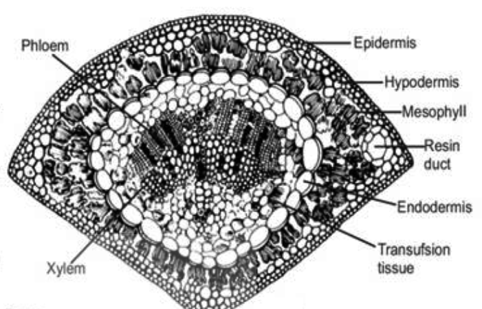
iii) T.S. needle of Pinus.
iv) L.S. microsporangium of Selaginella.
ii) V.S. thallus through a gemma cup in Marchantia.
iii) T.S. needle of Pinus.
iv) L.S. microsporangium of Selaginella.
Answer:
i) Clamp connection formation in Basidiomycetes.

ii) V.S. thallus through a gemma cup in Marchantia.

iii) T.S. needle of Pinus.
iv) L.S. microsporangium of Selaginella.

Question:-6(a)
Describe the internal and external structure of a typical bacterium.
Answer:
1. Overview of Bacterial Structure
Bacteria are single-celled prokaryotic organisms, meaning they lack a membrane-bound nucleus and other organelles found in eukaryotic cells. Their structure is relatively simple yet highly efficient, enabling them to thrive in diverse environments. A typical bacterium, such as Escherichia coli, has both external and internal components that work together to ensure survival, reproduction, and adaptation. The external structure provides protection and interaction with the environment, while the internal structure houses the machinery for cellular processes. This dual organization reflects bacteria’s ability to maintain functionality despite their microscopic size, typically ranging from 0.5 to 5 micrometers.
2. External Structure: The Protective Envelope
The external structure of a bacterium serves as its interface with the outside world, offering defense and structural integrity. It consists of several key layers and appendages.
The cell wall is a rigid layer made of peptidoglycan, a polymer of sugars and amino acids, which gives bacteria their shape (e.g., rod-shaped bacilli or spherical cocci) and protects against osmotic pressure. Gram-positive bacteria have a thick peptidoglycan layer, while Gram-negative bacteria have a thinner layer surrounded by an outer membrane containing lipopolysaccharides, which adds extra protection and contributes to pathogenicity.
Surrounding the cell wall in many bacteria is the capsule, a polysaccharide layer that enhances virulence by shielding the bacterium from the host immune system and aiding in attachment to surfaces. Some bacteria also have a looser, slimy glycocalyx for similar purposes.
The plasma membrane, a phospholipid bilayer beneath the cell wall, regulates the entry and exit of substances, maintaining homeostasis. It also contains proteins for transport and signaling.
External appendages like flagella, long whip-like structures, enable motility, while pili (hair-like projections) facilitate attachment to surfaces or other cells. Fimbriae, shorter than pili, assist in adhesion, such as during biofilm formation. Together, these components equip bacteria to interact with and survive in their surroundings.
3. Internal Structure: The Functional Core
Inside the bacterium lies a compact yet highly organized interior, lacking membrane-bound organelles but containing essential structures for life processes.
The cytoplasm is a gel-like matrix filling the cell, composed of water, enzymes, nutrients, and waste products. It serves as the site for metabolic reactions, such as glycolysis. Suspended within the cytoplasm is the nucleoid, an irregularly shaped region where the bacterial chromosome—a single, circular DNA molecule—resides. Unlike eukaryotic nuclei, the nucleoid is not enclosed by a membrane, but it efficiently manages genetic information for replication and protein synthesis.
Ribosomes, smaller than their eukaryotic counterparts (70S vs. 80S), are scattered throughout the cytoplasm. These molecular machines translate mRNA into proteins, critical for bacterial growth and function. Their abundance reflects the rapid reproduction rates of bacteria.
Many bacteria contain plasmids, small, circular DNA molecules separate from the chromosome. Plasmids often carry genes for antibiotic resistance or specialized functions, providing adaptability.
Inclusion bodies are storage granules within the cytoplasm, holding nutrients like glycogen, lipids, or polyphosphate for use during scarcity. Some bacteria also have gas vesicles or magnetosomes—specialized structures for buoyancy or orientation in magnetic fields, respectively—demonstrating internal diversity despite simplicity.
4. Coordination Between Internal and External Structures
The internal and external structures of a bacterium are intricately linked to support its life cycle. The plasma membrane, while technically external, acts as a bridge, facilitating communication between the cytoplasm and the outside via transport proteins and receptors. The cell wall and capsule protect the delicate internal components from environmental stress, such as desiccation or immune attack, while flagella and pili enable the bacterium to seek favorable conditions, indirectly supporting internal metabolism.
Internally, the nucleoid directs the synthesis of proteins (via ribosomes) that maintain the external structures, like enzymes for peptidoglycan assembly. Plasmids may encode toxins or adhesins that enhance external interactions, such as pathogenicity or biofilm formation. This coordination ensures bacteria can respond to challenges, from nutrient shortages to host defenses, showcasing their resilience.
Conclusion
In summary, a typical bacterium exemplifies efficiency in its structural design. Externally, the cell wall, capsule, and appendages provide protection, motility, and attachment, while internally, the cytoplasm, nucleoid, ribosomes, and plasmids drive metabolism, genetics, and adaptability. This interplay between external and internal features allows bacteria to colonize diverse niches, from soil to human guts, and even extreme environments like hot springs. Understanding bacterial structure not only highlights their biological significance but also informs medical and industrial applications, such as antibiotic development and biotechnology. Despite their simplicity, bacteria’s structural sophistication underscores their evolutionary success.
Question:-6(b)
Differentiate a bacterial cell from an archaeal cell.
Answer:
1. Overview of Bacterial and Archaeal Cells
Bacteria and archaea are both prokaryotic microorganisms, meaning they lack a membrane-bound nucleus and organelles typical of eukaryotic cells. Despite their superficial similarities, they represent distinct domains of life with significant differences in cell structure, biochemistry, and genetics. Bacteria are ubiquitous, thriving in diverse environments like soil, water, and human bodies, while archaea are often found in extreme conditions, such as hot springs, salt lakes, and deep-sea vents. These differences stem from evolutionary divergence, reflected in their cellular architecture and molecular machinery, which this response will explore in detail.
2. Cell Wall Composition
One of the most striking differences between bacterial and archaeal cells lies in their cell walls. Bacterial cell walls are primarily composed of peptidoglycan, a polymer of sugars (N-acetylglucosamine and N-acetylmuramic acid) cross-linked by peptide chains. This structure provides rigidity and determines Gram-positive (thick peptidoglycan) or Gram-negative (thin peptidoglycan plus an outer membrane) classification. The presence of peptidoglycan is a hallmark of bacteria and a target for antibiotics like penicillin.
In contrast, archaeal cell walls lack peptidoglycan entirely. Instead, they may contain pseudopeptidoglycan (with different sugar linkages), polysaccharides, or proteins, depending on the species. Some archaea, like methanogens, have a proteinaceous S-layer as their sole wall component, offering flexibility and protection in extreme environments. This absence of peptidoglycan makes archaea resistant to many antibacterial drugs, highlighting a key biochemical distinction.
3. Membrane Structure and Lipids
The plasma membrane of bacterial and archaeal cells differs fundamentally in lipid composition. Bacterial membranes consist of phospholipids with ester-linked fatty acid chains, forming a bilayer similar to eukaryotic membranes. This structure is relatively uniform across bacterial species, with variations in fatty acid saturation affecting fluidity.
Archaeal membranes, however, feature ether-linked lipids, often with branched isoprenoid chains instead of straight fatty acids. These ether linkages are more stable under extreme conditions like high temperature or acidity. In some archaea, such as thermophiles, the lipids form a monolayer rather than a bilayer, enhancing membrane durability. This adaptation allows archaea to survive in harsh habitats where bacteria typically cannot, marking a significant structural divergence.
4. Genetic and Molecular Machinery
Genetically, bacteria and archaea share the prokaryotic trait of a single, circular chromosome located in the nucleoid, but their molecular machinery reveals differences. Bacterial DNA replication, transcription, and translation rely on enzymes and processes distinct from eukaryotes. For example, bacterial RNA polymerase is simpler and sensitive to antibiotics like rifampicin.
Archaeal genetic machinery, conversely, resembles eukaryotic systems more closely. Their RNA polymerase and transcription factors are complex and similar to those in eukaryotes, and their replication proteins (e.g., DNA polymerase) share eukaryotic homologs. Archaeal ribosomes are 70S, like bacterial ones, but their sensitivity to inhibitors differs—archaea are resistant to many bacterial-specific drugs (e.g., chloramphenicol) and more akin to eukaryotic responses. This molecular hybridity underscores archaea’s unique evolutionary position.
5. Environmental Adaptations and Appendages
Bacterial and archaeal cells also differ in their environmental adaptations and surface structures. Bacteria often possess flagella and pili, protein-based appendages for motility and attachment, respectively. These structures are common across diverse bacterial species, aiding in colonization or pathogenesis. Capsules and glycocalyx layers further enhance bacterial survival in varied niches.
Archaeal flagella (termed archaella) differ structurally and functionally—they are thinner, powered by ATP rather than a proton motive force, and evolutionarily distinct from bacterial flagella. Pili-like structures exist in some archaea, but their roles are less studied. Archaea lack capsules in many cases, relying instead on their robust membranes and walls for protection in extreme conditions, such as high salinity or pH, where bacteria are less likely to thrive.
Conclusion
In summary, bacterial and archaeal cells, while both prokaryotic, diverge significantly in cell wall composition, membrane lipids, genetic machinery, and environmental adaptations. Bacteria’s peptidoglycan walls and ester-linked membranes suit them for diverse, often moderate environments, whereas archaea’s pseudopeptidoglycan or protein walls and ether-linked lipids equip them for extreme habitats. Genetically, archaea bridge bacteria and eukaryotes, with molecular systems reflecting this intermediary role. These distinctions not only highlight their evolutionary separation but also explain their ecological niches—bacteria as versatile generalists and archaea as extremophiles. Understanding these differences is crucial for microbiology, biotechnology, and astrobiology, revealing the remarkable diversity within prokaryotic life.
Question:-7(a)
Discuss different types of steles found in pteridophytes along with suitable diagrams.
Answer:
1. Introduction to Steles in Vascular Plants
In vascular plants, the vascular tissue (xylem and phloem) forms the stelar system, which plays a crucial role in water, nutrients, and food transport. The stelar theory is essential for understanding the structure of vascular cylinders in roots and stems. Based on this theory, scientists have identified various types of vascular arrangements, the two main categories being protostele and siphonostele. These structures evolve and differ in their complexity, depending on the plant’s evolutionary development.
2. Protostele: The Primitive Vascular System
Protostele represents one of the most fundamental stelar systems, where a solid core of xylem is encircled by phloem and lacks a pith. This type of vascular structure is considered to be the precursor to more complex steles found in higher plants. Protosteles are common in early vascular plants like Rhynia, and they continue to exist in many modern pteridophytes such as Selaginella, Lycopodium, and Lygodium.
This basic structure of protostele has further evolved into various subtypes, depending on the shape and organization of the xylem. Protostele serves as an essential developmental blueprint for understanding plant evolution.
3. Haplostele: Circular and Uniform
The haplostele is the simplest form of protostele. In this structure, the xylem core is solid and circular in cross-section, surrounded by a uniform layer of phloem. This simple arrangement is a characteristic feature of plants such as Selaginella kraussiana, S. chrysocaulos, and Lygodium. The term “haplostele” was introduced by Brebner in 1902 to describe this particular structure. It is one of the most basic forms of vascular tissue organization, demonstrating the simplest vascular development in pteridophytes.
4. Actinostele: Radiating Xylem Ridges
The actinostele type is characterized by a central xylem core that extends into radiating ridges. The xylem core in an actinostele is typically star-shaped or stellate, and it is surrounded by phloem. The phloem is not arranged in a uniform cylinder but is found in separate groups that alternate with the arms of the star-shaped xylem. This structure can be observed in plants such as Asteroxylon, Psilotum, and Lycopodium.
In certain actinosteles, the xylem may take the form of plate-like structures arranged in parallel, and the phloem is found in transverse bands or plates. This configuration is seen in the genus Lycopodium, where the vascular arrangement is termed as plectostele.
5. Siphonostele: The Evolutionary Advancement
Siphonostele represents a more advanced form of vascular system, in which the central area of the stele is filled with pith, and the vascular tissues (xylem and phloem) are arranged around the pith. The siphonostele can be further divided into two main types: ectophloic and amphiphloic.
- Ectophloic Siphonostele: In this type, the xylem lies closest to the pith, and the phloem surrounds the xylem externally, forming a layer around the vascular tissue. This arrangement is typical of species like Osmunda and Schizaea. The xylem core is the central structure, with phloem wrapping around it externally.
- Amphiphloic Siphonostele: Here, the phloem is located on both sides of the xylem core, with the pith still occupying the center. This type of stele can be found in the stem of plants such as Marsilea. The presence of phloem on both sides of the xylem marks an advanced form of vascular tissue arrangement in the evolutionary spectrum of plants.
6. Solenostele: Modified Siphonostele in Ferns
The solenostele is a modification of the siphonostele found in certain ferns, where the leaves are spaced out widely enough to allow for the formation of clear gaps between the vascular strands. These gaps, known as leaf gaps, do not overlap, which results in a characteristic horse-shoe-shaped xylem structure. This type of stele is more commonly seen in ferns that possess broader leaves with sufficient space between them.
7. Dictyostele: A Complex Siphonostele
When the leaf gaps become larger and overlap in some ferns, the vascular cylinder is divided into a tubular network of interconnected longitudinal strands. These strands, called meristeles, are separated by vertical strips of parenchymatous tissue, known as leaf gaps. The meristeles, as seen in cross-section, form a ring-like structure. This arrangement is termed dictyostele or dissected siphonostele. It is commonly found in ferns where the shoot axis is short, and leaves are inserted in close succession.
Each meristele in a dictyostele retains the structure of a protostele, making this a complex and highly specialized form of siphonostele.
8. Polystele: Multiple Vascular Cylinders
Polystele refers to the presence of multiple vascular bundles or steles in a plant axis, which is a unique feature of certain pteridophytes. In species like Selaginella, multiple steles are found within the same plant, allowing for enhanced vascular connectivity. This type of stele is a more advanced form of vascular structure, further adding complexity to plant evolution.


Conclusion
In summary, the stelar system in plants is a crucial element of their vascular anatomy, providing a structural basis for water, nutrient, and food transport. From the primitive protostele to the advanced siphonostele, these vascular arrangements have evolved over time to adapt to the diverse needs of plants. Understanding the types of steles—haplostele, actinostele, siphonostele, solenostele, dictyostele, and polystele—provides important insight into plant evolution and the complexity of their vascular systems. The study of these different stelar types, particularly in pteridophytes, underscores the ongoing adaptation of plants to their environments and highlights the evolutionary path that has led to the diversity of vascular plants we see today.
Question:-7(b)
Briefly describe the economic importance of Pteridophytes or Gymnosperms.
Answer:
1. Introduction to Pteridophytes and Gymnosperms
Pteridophytes and gymnosperms are two distinct groups of vascular plants that have played an important role in the evolution of plant life. Pteridophytes, also known as ferns and their relatives, include plants that reproduce via spores rather than seeds, while gymnosperms, or conifers, produce seeds that are not enclosed in a fruit. Both groups have contributed to the ecological stability of various habitats and have significant economic importance due to their medicinal, industrial, and ecological values. While pteridophytes are often overlooked in comparison to flowering plants, their contributions to human society and the environment are substantial. Gymnosperms, on the other hand, are more widely recognized for their role in forestry, agriculture, and their ability to adapt to a range of environmental conditions.
2. Economic Importance of Pteridophytes
Pteridophytes, despite being less common in economic discussions, have numerous practical uses in various industries. Their contribution can be categorized into several key areas:
- Medicinal Uses: Many species of ferns, such as Dryopteris filix-mas (male fern), have been traditionally used for medicinal purposes. Ferns are known to have anti-parasitic properties and have been utilized to treat intestinal worms. The extracts from certain ferns are also believed to have anti-inflammatory and anti-cancer properties, making them valuable in modern pharmaceutical research.
- Horticultural Uses: Pteridophytes are commonly used in the landscaping and horticulture industries. Ferns are popular ornamental plants due to their lush green appearance and their ability to thrive in shady areas. They are often used in indoor gardening, as well as in greenhouses and public parks. The aesthetic appeal of ferns, with their delicate fronds and diverse textures, makes them valuable in various design schemes, from home gardens to larger-scale landscaping projects.
- Soil Erosion Control: Ferns, particularly those found in damp, shaded environments, play an important role in preventing soil erosion. Their roots help bind the soil, preventing it from being washed away by heavy rainfall. This makes ferns an essential part of soil conservation efforts in areas prone to erosion, such as slopes or regions with poor vegetation cover.
- Ecological Significance: Pteridophytes are key components of the ecosystem, contributing to nutrient cycling and supporting a wide variety of wildlife. Many species of ferns provide habitats for small insects, amphibians, and birds. They also contribute to the stabilization of forest ecosystems by creating microhabitats and maintaining the balance of moisture and temperature.
- Cultural Significance: In many cultures, ferns have symbolic meanings and are used in traditional rituals and practices. For example, the fern is a symbol of new beginnings and protection in various cultures, particularly in New Zealand and other parts of the Pacific Islands. The fronds of certain ferns are also used in ceremonial arrangements and decorative arts.
3. Economic Importance of Gymnosperms
Gymnosperms, particularly conifers, have a much more prominent economic role compared to pteridophytes. These plants provide a wide range of resources used in industries ranging from timber production to pharmaceuticals. Below are some key areas in which gymnosperms contribute economically:
- Timber and Pulpwood: One of the most significant economic contributions of gymnosperms is their timber. Trees such as Pine, Spruce, and Fir are heavily harvested for their wood, which is used in construction, furniture making, and paper production. Gymnosperms are known for their straight trunks and high-quality wood, making them ideal for building materials. The wood of these trees is also processed into pulp for making paper, which is a multi-billion-dollar industry globally.
- Resins and Turpentine: Gymnosperms, particularly pines, are well known for producing resins. These resins are collected and used in the production of products such as turpentine, rosin, and adhesives. Turpentine is a valuable solvent used in paints, varnishes, and cleaning agents, while rosin is used in various industrial applications, including as a component in the production of rubber and as a lubricant in machinery.
- Medicinal Uses: Several gymnosperms have been studied for their medicinal properties. For example, Ginkgo biloba, a species of gymnosperm, is widely used in traditional medicine and has been shown to have cognitive-enhancing properties. It is particularly used in the treatment of memory loss and other cognitive disorders, making it an important plant in the pharmaceutical industry.
- Ecological Significance: Gymnosperms, particularly in temperate regions, are essential for stabilizing ecosystems. They are adapted to survive in harsh climates, including cold, dry, and nutrient-poor soils, which allows them to play a crucial role in maintaining biodiversity in these regions. Coniferous forests are critical habitats for many species of wildlife, including birds, mammals, and insects. The ecological role of gymnosperms in carbon sequestration also contributes to their importance in the fight against climate change.
- Ornamental Uses: Many species of gymnosperms, such as Christmas trees (e.g., Fir, Pine, and Spruce), are economically important as ornamental plants. These trees are in high demand during the holiday season, contributing to a significant seasonal industry. Additionally, coniferous shrubs and trees are used in landscaping and as hedging plants due to their attractive appearance, hardiness, and evergreen nature.
- Food Products: Certain species of gymnosperms, such as Pine nuts from pine trees, are harvested for food. Pine nuts are a popular ingredient in many dishes, including Mediterranean and Asian cuisines. They are also used to make pine nut oil, which is highly valued in culinary and cosmetic industries for its health benefits and versatile uses.
4. Challenges and Sustainability of Pteridophytes and Gymnosperms
While both pteridophytes and gymnosperms have significant economic value, they face a range of challenges related to sustainability and environmental impact. Overharvesting of certain species for their timber, resins, and medicinal properties can lead to population declines and, in some cases, the risk of extinction. Deforestation, habitat loss, and climate change further exacerbate these issues. In response, there is an increasing push for sustainable harvesting practices, reforestation efforts, and the development of alternative materials to reduce the pressure on these plant groups.
For pteridophytes, conservation efforts are focused on preserving their natural habitats, especially in areas where their role in soil stabilization and biodiversity is critical. Gymnosperms are also benefiting from initiatives that promote sustainable forestry, including certification systems such as the Forest Stewardship Council (FSC), which ensures that timber is sourced from responsibly managed forests.
5. Conclusion
Pteridophytes and gymnosperms are two vital groups of plants that contribute significantly to the economy, ecology, and human well-being. Pteridophytes, though often overlooked, provide essential services in horticulture, soil conservation, and medicine. On the other hand, gymnosperms have a more prominent role in industries such as timber, resin production, and pharmaceuticals. Both plant groups have unique characteristics that make them invaluable resources, but their long-term sustainability requires careful management and conservation efforts. As we continue to rely on these plants for various economic, environmental, and medicinal benefits, it is essential to ensure that their harvest and use are done in a way that does not threaten their future existence. Sustainable practices and policies are key to preserving the value of pteridophytes and gymnosperms for future generations.
Question:-8
Prepare a detailed account of occurrence, morphology and ultrastructure in Cyanophyta.
Answer:
1. Introduction to Cyanophyta
Cyanophyta, commonly known as blue-green algae, are a group of photosynthetic organisms that play a crucial role in the environment and ecology of aquatic ecosystems. They are a part of the Prokaryotae (or Prokaryotes) domain and are one of the oldest and most diverse groups of organisms on Earth. They are unique due to their ability to perform oxygenic photosynthesis, similar to plants. The term Cyanophyta refers to their characteristic blue-green pigment, phycocyanin, which gives them their distinctive color. Cyanophytes can be found in a wide range of habitats, including freshwater, marine, and terrestrial environments. Their occurrence and morphology are of significant interest, as they have been pivotal in shaping life on Earth for billions of years.
2. Occurrence of Cyanophyta
Cyanophytes are widely distributed in nature, occurring in diverse environments ranging from freshwater lakes, ponds, and rivers to marine environments. They can also be found in extreme habitats like hot springs, deserts, and even in symbiotic relationships with other organisms such as lichens. Cyanophytes are particularly abundant in nutrient-rich waters where they can proliferate quickly, forming blooms that can impact water quality and ecology. The global distribution of Cyanophyta is driven by their ability to adapt to various environmental conditions, including variations in temperature, pH, and salinity. Some species are capable of surviving extreme conditions, such as very high or low temperatures, making them one of the most versatile groups of organisms on Earth.
The ability of Cyanophyta to thrive in both oligotrophic (low nutrient) and eutrophic (high nutrient) waters makes them a key component in aquatic ecosystems. Their presence in aquatic habitats plays a crucial role in maintaining the balance of the ecosystem, especially through nitrogen fixation, a process where they convert atmospheric nitrogen into a form usable by plants. This process contributes to the overall fertility of aquatic environments, supporting a variety of other organisms.
3. Morphology of Cyanophyta
Cyanophyta exhibit a wide range of morphological forms. Their structure is relatively simple compared to other algae and plants, which is typical of prokaryotic organisms. Cyanophytes can exist as unicellular organisms, colonial forms, or filamentous forms.
- Unicellular Forms: Many cyanophytes exist as single-celled organisms. These cells are generally spherical, oval, or rod-shaped. In unicellular species, the entire cell is responsible for photosynthesis, and the cyanophyte can move through water via gliding motility. Unicellular cyanophytes include Microcystis and Synechococcus.
- Colonial Forms: Some cyanophytes form aggregates or colonies of cells that work together. These colonies can be composed of hundreds to thousands of individual cells. An example of a colonial cyanophyte is Merismopedia, which forms flat, sheet-like colonies.
- Filamentous Forms: Certain cyanophytes form long chains or filaments, where individual cells are linked together by a mucilaginous sheath. Filamentous species are often more complex, with some specialized cells performing specific functions. Nostoc and Anabaena are examples of filamentous cyanophytes. These organisms may also form specialized cells such as heterocysts, which are involved in nitrogen fixation.
Cyanophytes are often surrounded by a gelatinous sheath or mucilage, which provides protection and helps them to adhere to surfaces or form colonies. This mucilage is an important feature of many cyanophytes and helps in their survival in variable environmental conditions. The cells contain chlorophyll a and phycocyanin, pigments that allow them to absorb light and carry out photosynthesis.
4. Ultrastructure of Cyanophyta
The ultrastructure of cyanophytes reveals a relatively simple but highly effective organization of cellular components, characteristic of prokaryotic organisms. Unlike eukaryotic cells, cyanophytes do not have membrane-bound organelles such as a nucleus or mitochondria. However, they possess specialized structures that perform the functions necessary for survival and reproduction.
- Cell Membrane: The plasma membrane in cyanophytes is surrounded by a rigid cell wall made of peptidoglycan, which provides structural support and protects the cell. The membrane contains photosynthetic enzymes and protein complexes essential for photosynthesis.
- Thylakoids: Instead of chloroplasts, cyanophytes contain thylakoid membranes, which are flattened sacs containing the photosynthetic pigments (chlorophyll a and phycocyanin). These thylakoids are organized into layers within the cytoplasm and are the sites where photosynthesis takes place. Cyanophytes are able to perform oxygenic photosynthesis using sunlight, carbon dioxide, and water.
- Nucleoid: The genetic material of cyanophytes is found in a region called the nucleoid, which is not enclosed by a membrane. The nucleoid contains a single, circular chromosome of DNA, which carries the genetic information for the cell. Cyanophytes may also contain small, circular DNA molecules known as plasmids, which carry additional genetic material.
- Gas Vesicles: Many cyanophytes contain gas vesicles, which are protein-bound structures filled with gas that help the organisms maintain buoyancy. These vesicles allow cyanophytes to move toward or away from light, a process known as phototaxis, which optimizes their exposure to sunlight for photosynthesis.
- Heterocysts: Some cyanophytes, especially filamentous forms like Anabaena, form specialized cells called heterocysts. These cells are involved in nitrogen fixation, where atmospheric nitrogen (N2) is converted into ammonia, a form that can be used by plants. Heterocysts are characterized by their thickened cell walls, which protect the nitrogenase enzyme responsible for nitrogen fixation from oxygen, which can inhibit its activity.
- Reproduction: Cyanophytes primarily reproduce asexually through binary fission, where a single cell divides into two identical daughter cells. Some cyanophytes, particularly filamentous species, may also reproduce through the formation of akinetes, which are specialized resting cells that can survive unfavorable conditions and germinate when conditions improve.
5. Ecological Significance of Cyanophyta
Cyanophytes play a critical role in the environment, particularly in aquatic ecosystems. As primary producers, they form the base of the food chain in many aquatic habitats, providing essential nutrients to a wide variety of organisms, from small zooplankton to larger fish. Their ability to fix nitrogen also enhances the productivity of ecosystems by making nitrogen available to other organisms.
In some environments, cyanophytes can form harmful algal blooms, which can have detrimental effects on water quality. These blooms can deplete oxygen levels, leading to hypoxia, and produce toxins that can harm aquatic life and humans. Despite these occasional negative impacts, cyanophytes are overall vital components of ecosystems.
Conclusion
Cyanophyta are ancient organisms that have evolved to occupy diverse habitats, ranging from freshwater lakes to extreme environments. Their morphology varies from unicellular to filamentous forms, and they play vital roles in ecosystems, including nitrogen fixation and serving as primary producers. The ultrastructure of cyanophytes reflects their prokaryotic nature, with specialized structures such as thylakoids for photosynthesis, gas vesicles for buoyancy, and heterocysts for nitrogen fixation. These unique features allow cyanophytes to thrive in a wide range of conditions, contributing to the health of aquatic environments and offering potential applications in biotechnology, agriculture, and environmental management.
Question:-9
Define the transformation. How Griffith’s experiment was carried out to discover bacterial transformation?
Answer:
1. Definition of Transformation
Transformation is a process in molecular biology where a cell takes up foreign genetic material (usually DNA) from its environment and incorporates it into its own genome. This process can lead to the genetic modification of the cell, which can result in changes in its phenotype. In bacteria, transformation occurs naturally in some species, allowing them to adapt to environmental changes. The process of bacterial transformation has been a significant tool in genetics, molecular biology, and biotechnology, as it allows for the manipulation of genes and the introduction of new genetic traits into organisms.
Transformation was first observed in bacteria by Frederick Griffith in 1928, and it became one of the foundational discoveries in the field of genetics. This phenomenon demonstrated that genetic material could be transferred between bacterial cells, even without direct cell-to-cell contact, highlighting the role of DNA in heredity and genetic change.
2. Griffith’s Experiment: Discovery of Bacterial Transformation
Frederick Griffith’s experiment, conducted in 1928, was a pivotal study in understanding bacterial genetics. The purpose of the experiment was to explore the nature of bacterial pneumonia caused by Streptococcus pneumoniae. Griffith was particularly interested in how different strains of the bacterium might behave and how they could cause disease.
Griffith worked with two distinct strains of Streptococcus pneumoniae: a virulent (disease-causing) strain and a non-virulent (harmless) strain. The key difference between the two strains was the presence or absence of a smooth, protective capsule surrounding the bacterial cell. The smooth strain (S strain) had a capsule and was virulent, capable of causing pneumonia, while the rough strain (R strain) lacked a capsule and was non-virulent, meaning it was unable to cause disease.
3. Griffith’s Experimental Setup
Griffith carried out a series of injections using the two strains of bacteria into mice to test their effects on the health of the animals. The experiment involved four key groups:
- Injection of the S strain (virulent strain): When Griffith injected the mice with the S strain, they developed pneumonia and died, as expected because the S strain was capable of causing disease due to the protective capsule.
- Injection of the R strain (non-virulent strain): When the mice were injected with the R strain, they remained healthy, as the R strain was not able to cause disease due to the absence of a protective capsule.
- Injection of heat-killed S strain: Griffith then heated the S strain bacteria to kill them, and injected the heat-killed S strain into the mice. The mice survived, as the dead bacteria could not cause disease.
- Injection of heat-killed S strain mixed with live R strain: In the most surprising part of the experiment, Griffith injected mice with a mixture of heat-killed S strain and live R strain. To his surprise, the mice died from pneumonia, and live S strain bacteria were found in their blood.
4. Observations and Conclusion
Griffith’s observation of the mice that were injected with the mixture of heat-killed S strain and live R strain led to the discovery of bacterial transformation. When the heat-killed S strain and live R strain were mixed, some of the R strain bacteria acquired the genetic material from the dead S strain, and transformed into virulent S strain bacteria. These transformed bacteria were capable of producing a capsule, which made them virulent and able to cause disease.
Griffith concluded that some "transforming principle" was passed from the dead S strain to the live R strain, causing the R strain to acquire the ability to make a capsule and become virulent. This was the first experimental evidence suggesting that genetic information could be transferred between bacteria, even without direct cell-to-cell contact.
5. Significance of Griffith’s Discovery
Griffith’s experiment laid the foundation for the discovery of DNA as the genetic material. At the time, the biochemical nature of the "transforming principle" was unknown, and many scientists believed proteins were the hereditary material. Griffith’s discovery prompted further research into the nature of transformation and, eventually, led to the identification of DNA as the molecule responsible for carrying genetic information.
The idea that bacteria could exchange genetic material through transformation opened the door to the development of genetic engineering techniques and modern molecular biology. It was later shown that the "transforming principle" was, in fact, DNA, a discovery confirmed by subsequent experiments, including the famous work of Avery, MacLeod, and McCarty in 1944.
6. Implications of Griffith’s Experiment in Modern Genetics
Griffith’s discovery of transformation has had far-reaching implications in molecular biology and biotechnology. Understanding transformation has allowed scientists to manipulate bacterial genetics, leading to advances in areas such as:
- Genetic Engineering: Transformation is a cornerstone technique in genetic engineering, where foreign genes are introduced into organisms (such as bacteria or plants) to modify their genetic makeup for practical purposes, including the production of proteins, vaccines, and other valuable compounds.
- Gene Therapy: Bacterial transformation has inspired the development of gene therapy techniques, where genetic material is introduced into human cells to treat genetic disorders.
- Antibiotic Resistance: Transformation is also an important mechanism by which bacteria can acquire antibiotic resistance genes from other bacteria. This has significant implications for public health, as it helps explain how bacterial populations evolve resistance to drugs.
Conclusion
Griffith’s experiment in 1928 was a landmark study in genetics, as it provided the first evidence of bacterial transformation. His work demonstrated that genetic material could be transferred between bacteria, allowing non-virulent strains to acquire virulence factors. This discovery was crucial in advancing our understanding of genetics and molecular biology, eventually leading to the identification of DNA as the genetic material. Griffith’s experiment laid the groundwork for modern genetic engineering techniques, biotechnology, and gene therapy, which have revolutionized medicine and science. The concept of transformation continues to be a powerful tool for researchers, with implications for medicine, agriculture, and environmental science.
Question:-10
Write notes on the following:
i) Biofertilizer
ii) Polyembryony in gymnosperms
iii) Alternation of generations
iv) Coralloid roots of Cycas
ii) Polyembryony in gymnosperms
iii) Alternation of generations
iv) Coralloid roots of Cycas
Answer:
i) Biofertilizer
Biofertilizers are natural fertilizers that consist of living microorganisms, which enhance the nutrient content of the soil, thereby promoting the growth of plants. These microorganisms include bacteria, fungi, algae, and actinomycetes, which are capable of fixing nitrogen, decomposing organic material, or solubilizing nutrients like phosphorus and potassium in the soil. Biofertilizers are considered a sustainable and eco-friendly alternative to chemical fertilizers, as they do not cause soil degradation or water pollution, unlike synthetic fertilizers. They are often used in combination with other organic farming practices to improve soil health and increase agricultural productivity.
Common examples of biofertilizers include Rhizobium, which fixes nitrogen in leguminous plants, Azotobacter and Azospirillum for nitrogen fixation in non-legumes, and Mycorrhizal fungi, which form symbiotic relationships with plant roots to help in the uptake of nutrients like phosphorus. Phosphate-solubilizing bacteria and blue-green algae (such as Anabaena and Nostoc) are also widely used to improve soil fertility.
The use of biofertilizers is growing due to their benefits in enhancing soil fertility, improving crop yields, and reducing the reliance on chemical fertilizers. Moreover, biofertilizers contribute to soil conservation and have a minimal environmental impact, promoting sustainable agriculture and ensuring food security for the future. Their application requires careful consideration of environmental conditions, as their efficacy can depend on factors like soil temperature, moisture, and pH.
ii) Polyembryony in Gymnosperms
Polyembryony is a phenomenon where multiple embryos develop from a single fertilized egg cell, leading to the formation of multiple seedlings from a single ovule. This process occurs in various plant species, including gymnosperms such as Cycas, Pinus, and Ginkgo. In gymnosperms, polyembryony can result from the development of more than one embryo from the same fertilized ovule, typically due to the presence of additional embryos formed from the same megaspore mother cell or the fertilization of multiple egg cells.
In gymnosperms, polyembryony often occurs in species like Pinus and Cycas, where more than one embryo is produced from a single ovule. The phenomenon is particularly well-known in Cycas, where the embryos may arise from the megaspore or from the surrounding tissues, including the nucellus. The presence of multiple embryos can be beneficial for the plant’s survival, as it increases the chances of successful germination and establishment in varying environmental conditions.
Polyembryony in gymnosperms is considered a result of a genetic or epigenetic event and is useful in horticulture and plant breeding. For instance, the extra embryos can be selected and grown into independent plants, providing opportunities for clonal propagation. Furthermore, polyembryony helps in the conservation of rare or endangered gymnosperm species by producing multiple genetically identical plants from a single seed.
iii) Alternation of Generations
Alternation of generations is a biological cycle in which organisms alternate between two distinct multicellular stages: a diploid sporophyte generation and a haploid gametophyte generation. This phenomenon is observed in plants, algae, and some fungi. The sporophyte generation produces spores through meiosis, which are haploid and can grow into gametophytes. The gametophyte then produces gametes (sperm and egg cells) by mitosis, which fuse during fertilization to form a diploid zygote. The zygote then grows into the next sporophyte generation, completing the cycle.
In plants, this alternation occurs between the sporophyte (the diploid, spore-producing plant) and the gametophyte (the haploid, gamete-producing structure). In vascular plants like ferns, the sporophyte is the dominant, visible form, while the gametophyte is small and independent. In non-vascular plants (such as mosses), the gametophyte is the dominant phase, and the sporophyte is dependent on it for nourishment.
Alternation of generations allows for genetic diversity through the reshuffling of genes during meiosis and fertilization. This process also enables plants to adapt to varying environmental conditions, as it combines the advantages of both sexual and asexual reproduction. For example, while the sporophyte generation can disperse spores over long distances, the gametophyte generation ensures genetic variability through sexual reproduction.
iv) Coralloid Roots of Cycas
Coralloid roots are specialized roots found in certain plants, particularly in the cycad family, such as Cycas species. These roots are unique because of their structure, which resembles coral, hence the name “coralloid.” They are typically found growing above ground and are characterized by their branched, fleshy, and nodular appearance. The function of coralloid roots is closely linked to their symbiotic relationship with nitrogen-fixing cyanobacteria, such as Nostoc and Anabaena, which colonize the roots and convert atmospheric nitrogen into ammonia, a form that the plant can utilize for growth.
In Cycas, the coralloid roots grow in clusters and have specialized structures called cephalodiums that house the cyanobacteria. These bacteria provide the plant with a continuous supply of nitrogen, which is especially beneficial for plants growing in nitrogen-poor soils. The plant provides a suitable environment for the cyanobacteria, including moisture and nutrients, forming a mutualistic relationship.
The coralloid roots also help in the overall nutrient uptake and growth of the plant. By hosting these bacteria, Cycas plants can survive in environments where other plants might struggle due to the low availability of nitrogen. This adaptation has made cycads like Cycas particularly well-suited to their habitats, allowing them to thrive in arid and nutrient-poor conditions. The study of coralloid roots provides insights into the fascinating symbiotic relationships that exist in nature, as well as the evolution of plant adaptation mechanisms.